


Gonarthrosisis an arthrosis deformity of the knee joint. It is accompanied by damage to the hyaline cartilage of the articular surface of the tibia and femur and has a chronic progressive course. Clinical symptoms include pain that worsens with movement, limitation of movement, and synovitis (accumulation of fluid) in the joint. At a later stage, the support on the legs is affected, and a clear limitation of movement is observed. Pathology is diagnosed based on anamnesis, complaints, physical examination and joint radiography. Treatment is conservative: drug therapy, physiotherapy, exercise therapy. If there is significant destruction of the joint, endoprosthetics are indicated.
General information
Gonarthrosis (from the Latin genus articulatio - knee joint) or deformed knee joint arthrosis is a progressive degenerative-dystrophic lesion of intra-articular cartilage that is not inflammatory in nature. Gonarthrosis is the most common arthrosis. Usually affects middle-aged and elderly people, women are more often affected. After injury or continuous intense stress (for example, during professional sports), gonarthrosis can occur at a younger age. Prevention plays the most important role in preventing the occurrence and development of gonarthrosis.
Contrary to popular belief, the cause of the development of this disease does not lie in the deposition of salt, but in malnutrition and changes in the structure of intra-articular cartilage. With gonarthrosis, a concentration of calcium salt deposition may occur at the site of attachment of tendons and ligament apparatus, but it is secondary and does not cause painful symptoms.
Causes of gonarthrosis
In most cases, it is impossible to identify any single reason for the development of pathology. As a rule, the occurrence of gonarthrosis is caused by a combination of several factors, including:
- injury. About 20-30% of gonarthrosis cases are associated with previous injuries: tibia fractures (especially intra-articular), meniscus injuries, ligament tears or ruptures. Usually, gonarthrosis occurs 3-5 years after a traumatic injury, although the early development of the disease is possible - 2-3 months after the injury.
- Physical training. Often the manifestation of gonarthrosis is associated with excessive load on the joints. The age after 40 is a period where many people understand that regular physical activity is necessary to keep the body in good condition. When starting to exercise, they do not take into account age-related changes and do not need to load the joints, which leads to the rapid development of degenerative changes and the appearance of gonarthrosis symptoms. Intense fast running and squatting are especially dangerous for the knee joint.
- Overweight. With excess body weight, the load on the joints increases, both microtrauma and serious damage (meniscus tears or ligament tears) occur more often. Gonarthrosis is particularly difficult in obese patients with severe varicose veins.
The risk of gonarthrosis also increases after previous arthritis (psoriatic arthritis, reactive arthritis, rheumatoid arthritis, gouty arthritis or ankylosing spondylitis). In addition, risk factors for the development of gonarthrosis include genetically determined weaknesses in the ligament apparatus, metabolic disorders and conservation disorders in certain neurological diseases, traumatic brain injuries and spinal injuries.
Pathogenesis
The knee joint is formed by the articular surfaces of two bones: the femur and the tibia. On the front surface of the joint is the patella, which, when moving, slides along the depression between the condyles of the femur. The fibula does not participate in the formation of the knee joint. Its upper part is located on the side and just below the knee joint and is connected to the tibia through a joint that moves low.
The articular surface of the tibia and femur, as well as the posterior surface of the patella, is covered with smooth, very strong and elastic dense hyaline cartilage 5-6 mm thick. Cartilage reduces frictional forces during movement and performs shock absorbing functions during shock loads.
In the first stage of gonarthrosis, blood circulation in the small intraosseous vessels that feed the hyaline cartilage is disrupted. The cartilage surface becomes dry and gradually loses its smoothness. Cracks are visible on the surface. Instead of sliding smoothly and without obstacles, the cartilage "clings" to each other. Due to continuous microtrauma, cartilage tissue becomes thinner and loses its shock-absorbing properties.
In the second stage of gonarthrosis, compensatory changes occur in the bone structure. The joint platform is flattened, adapting to the increased load. The subchondral zone (the part of the bone that lies just below the cartilage) is thickened. Bone growths appear along the edges of the articular surface - osteophytes, which in their appearance on radiographs resemble thorns.
During gonarthrosis, the synovial membrane and joint capsule also deteriorate and become "wrinkled. "The nature of the joint fluid changes - it thickens, its viscosity increases, which leads to a deterioration in its lubricating and nutritional properties. Due to the lack of nutrients, cartilage degeneration accelerates. Cartilage becomes thinner and in some areas disappears completely. After the loss of cartilage, the friction between the articular surfaces increases dramatically, and degenerative changes rapidly develop.
In the third stage of gonarthrosis, the bones are significantly deformed and seem to be pressed against each other, significantly limiting movement in the joint. Cartilaginous tissue is practically absent.
Classification
Taking into account the pathogenesis in traumatology and orthopedics, two types of gonarthrosis are distinguished: primary (idiopathic) and secondary gonarthrosis. Primary gonarthrosis occurs without previous trauma in elderly patients and is usually bilateral. Secondary gonarthrosis develops against the background of pathological changes (diseases, developmental disorders) or injuries of the knee joint. Can occur at any age, usually unilateral.
Taking into account the severity of pathological changes, three stages of gonarthrosis are distinguished:
- First stage- early manifestations of gonarthrosis. Characterized by periodic dull pain, usually after a significant load on the joint. There may be some swelling in the joint that goes away on its own. No deformation.
- Second stage– increased symptoms of gonarthrosis. The pain becomes longer and more intense. Buzzing sounds often appear. There is little or moderate movement restriction and little joint deformity.
- Third stage– clinical manifestations of gonarthrosis reach a maximum. The pain is almost constant, the gait is disturbed. There is significant mobility limitation and significant joint deformity.
Symptoms of gonarthrosis
The disease begins gradually, gradually. In the first stage of gonarthrosis, the patient experiences minor pain when moving, especially when going up or down stairs. There may be a feeling of stiffness in the joints and "tightening" in the popliteal area. A characteristic symptom of gonarthrosis is "starting pain" - a painful sensation that occurs during the first step after rising from a sitting position. When patients with gonarthrosis "diverge, " the pain decreases or disappears, and after significant pressure it reappears.
Externally the knee does not change. Sometimes patients with gonarthrosis note a slight swelling in the affected area. In some cases, in the first stage of gonarthrosis, fluid accumulates in the joint - synovitis develops, which is characterized by an increase in the volume of the joint (it becomes swollen, spherical), a feeling of heaviness and limitation of movement.
In the second stage of gonarthrosis, the pain becomes stronger, occurs even with a light load and increases with intense or long walking. As a rule, the pain is localized along the anterior inner surface of the joint. After a long rest, the painful sensation usually disappears, and reappears with movement.
As gonarthrosis develops, the range of motion in the joint gradually decreases, and when trying to bend the leg as much as possible, a sharp pain appears. There may be a rough pulsing sound when moving. The configuration of the joint changes, as if it is growing. Synovitis appears more often than in the first stage of gonarthrosis and is characterized by a more continuous course and accumulation of more fluid.
In the third stage of gonarthrosis, the pain becomes almost constant, bothering the patient not only while walking, but also at rest. At night, patients spend a long time trying to find a comfortable position to sleep. Often the pain appears even at night.
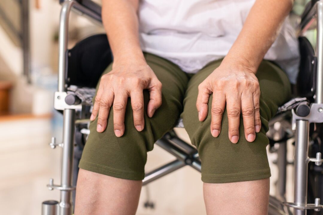
Flexion at the joint is significantly limited. In some cases, not only flexion, but also extension is limited, which is why patients with gonarthrosis cannot fully straighten their legs. Joints are enlarged and deformed. Some patients suffer from hallux valgus or varus deformation - the foot becomes X or O shaped. Due to the limited movement and deformation of the foot, the gait becomes unstable and wobbly. In severe cases, patients with gonarthrosis can move only with the support of crutches or crutches.
Diagnostics
The diagnosis of gonarthrosis is made based on the patient's complaints, objective examination data and x-ray examination. When examining patients with the first stage of gonarthrosis, external changes are usually not detected. In the second and third stages of gonarthrosis, the roughness of bone contours, joint deformation, limitation of movement and curvature of the axis of the limb are detected. When the patella moves in a transverse direction, a pulsating sound is heard. Palpation reveals painful areas inward from the patella, at the level of the joint space, as well as above and below it.
With synovitis, the joint increases in volume, its contour becomes smooth. A bulge is detected along the anterolateral surface of the joint and above the patella. After palpation, fluctuations are determined.
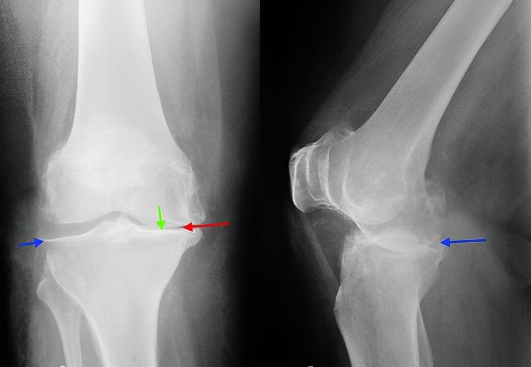
X-ray of the knee joint is a classic technique that allows you to clarify the diagnosis, establish the severity of pathological changes in gonarthrosis and monitor the dynamics of the process, taking repeated pictures after some time. Due to its availability and low cost, it remains the main method for diagnosing gonarthrosis to this day. In addition, this research method allows us to exclude other pathological processes (for example, tumors) in the tibia and femur.
In the early stages of gonarthrosis, changes on radiographs may not be present. After that, narrowing of the joint space and compaction of the subchondral zone are determined. The articular end of the femur and especially the tibia expands, the edge of the condyle becomes pointed.
When studying radiographs, it should be taken into account that more or less significant changes characteristic of gonarthrosis are observed in most elderly people and are not always accompanied by pathological symptoms. The diagnosis of gonarthrosis is made only by a combination of radiological and clinical signs of the disease.
Currently, along with traditional radiography, modern techniques such as computed tomography of the knee joint, which allows a detailed study of pathological changes in the bone structure, and MRI of the knee joint, which is used to identify changes in soft tissue, are used to diagnose gonarthrosis. .
Treatment of gonarthrosis
Conservative activity
The treatment is carried out by traumatologists and orthopedics. Therapy for gonarthrosis should begin as early as possible. During the exacerbation period, patients with gonarthrosis are recommended to rest for maximum unloading of the joint. Patients are prescribed therapeutic exercises, massage, physiotherapy (UHF, electrophoresis with novocaine, phonophoresis with hydrocortisone, diadynamic current, magnetic and laser therapy) and mud therapy.
Drug therapy for gonarthrosis includes chondroprotectors (drugs that increase metabolic processes in the joints) and drugs that replace synovial fluid. In some cases, with gonathrosis, intra-articular administration of steroid hormones is indicated. After that, the patient can be referred for sanatorium treatment.
Gonarthrosis patients may be advised to walk with crutches to unload the joint. Sometimes special orthoses or custom insoles are used. To slow down the degenerative process in the joints with gonarthrosis, it is very important to follow certain rules: exercise, avoid unnecessary stress on the joints, choose comfortable shoes, monitor your weight, organize your daily routine correctly (alternating loads and rest, doing special training).
Surgery
With obvious destructive changes (at the third stage of gonarthrosis), conservative treatment is ineffective. In cases of severe pain, joint dysfunction and limited ability to work, especially if young or middle-aged patients suffer from gonarthrosis, they resort to surgery (knee replacement). After that, recovery measures are carried out. The complete recovery period after joint replacement surgery for gonarthrosis takes from 3 months to six months.